

工作时间 :
周一~~周五
9:00 -18:00
在非工作时间,您可以通过邮件订购产品,订购时请写明详细联系方式,谢谢支持!
销售:18321282235
技术:021-60514606
传真:021-37680378


顾经理微信 扫一扫,关注我们

巨噬细胞炎性蛋白1α(MIP1a)活性蛋白: The thermal stability is described by the loss rate. The loss rate was determined by accelerated thermal degradation test, that is, incubate the protein at 37 oC for 48h, and no obvious degradation and precipitation were observe
[ PROPERTIES ]
Source: Eukaryotic expression.
Host: 293F cell
Residues: Ser24~Ala92
Tags: N-terminal His-tag
Purity: >95%
Endotoxin Level: <1.0EU per 1μg (determined by the LAL method).
Buffer Formulation: 20mM Tris, 150mM NaCl, pH8.0, containing 1mM EDTA, 1mM DTT, 0.01% sarcosyl, 5% trehalose, and Proclin300.
Applications: Cell culture; Activity Assays; In vivo assays.
(May be suitable for use in other assays to be determined by the end user.)
Predicted isoelectric point: 4.8
Predicted Molecular Mass: 9.3kDa
Accurate Molecular Mass: 14kDa as determined by SDS-PAGE reducing conditions.
Phenomenon explanation:
The possible reasons that the actual band size differs from the predicted are as follows:
1. Splice variants: Alternative splicing may create different sized proteins from the same gene.
2. Relative charge: The composition of amino acids may affects the charge of the protein.
3. Post-translational modification: Phosphorylation, glycosylation, methylation etc.
4. Post-translation cleavage: Many proteins are synthesized as pro-proteins, and then cleaved to give the active form.
5. Polymerization of the target protein: Dimerization, multimerization etc.
[ USAGE ]
Reconstitute in 20mM Tris, 150mM NaCl (pH8.0) to a concentration of 0.1-1.0 mg/mL. Do not vortex.
[ STORAGE AND STABILITY ]
Storage: Avoid repeated freeze/thaw cycles.
Store at 2-8C for one month.
Aliquot and store at -80for 12 months.
Stability Test: The thermal stability is described by the loss rate. The loss rate was determined by accelerated thermal degradation test, that is, incubate the protein at 37C for 48h, and no obvious degradation and precipitation were observed.The loss rate is less than 5% within the expiration date under appropriate storage condition.
[ SEQUENCE ] [ ACTIVITY ]
MIP-1a (macrophage inflammatory protein 1-alpha) also known as Chemokine (C-C motief) ligand 3 (CCL3), is a cytokine belonging to the CC chemokine family that is involved in the recruitment and activation of macrophages, monocytes and neutrophils. In this case, chemotaxis assay used 24-well microchemotaxis system was undertaken to evaluate the chemotactic effect of MIP-1a on the human monocytic cell line THP1. Briefly, THP1 cells were seeded into the upper chambers (100µl cell suspension, 106 cells/ml in RPMI 1640 with 0.5% FBS) and MIP-1a (100ng/mL, diluted in serum free RPMI 1640 ) was added in lower chamber with a polycarbonate filter (8µm pore size) used to separate the two compartments. After incubation at 37oC with 5% CO2 for 5h, the filter was removed, then cells in low chamber were observed by inverted microscope at low magnification (×40) and the number of migrated cells were counted at high
magnification (×400) randomly (five fields for each filter).
By counting migrated cells in low chamber at high magnification (×400) randomly, it was shown that a mean of 41.2 THP1 cells/field migrated towards serum free RPMI 1640 medium with 100ng/mL MIP-1a, while only 3.6 THP1 cells/field migrated towards serum free RPMI 1640 medium. And the migrated THP1 cells in low chamber at low magnification (×40) was shown in Figure 1.
Figure 1. The chemotactic effect of MIP-1-alpha on THP1 cells
(A) THP1 cells were seeded into the upper chambers and serum free RPMI 1640 with 100ng/mL MIP-1a was added in lower chamber, then cells in lower chamber were observed at low magnification (×40) after incubation for 5h;
(B) THP1 cells were seeded into the upper chambers and serum free RPMI 1640 with no MIP-1a was added in lower chamber, then cells in lower chamber were observed at low magnification (×40) after incubation for 5h.
[ IDENTIFICATION ]
Figure 2. Gene Sequencing (extract)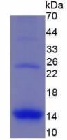
Figure 3. SDS-PAGE
Sample: Active recombinant MIP1a, Human
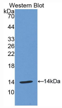
Figure 4. Western Blot
Sample: Recombinant MIP1a, Human;
Antibody: Rabbit Anti-Human MIP1a Ab (PAA092Hu06)
您可以点击 ![]() 下载产品说明书, 或直接联系我们订购.
下载产品说明书, 或直接联系我们订购.
部门 |
姓名 | 手机 | |||
| 销售部 | 顾先生 | 1916510334@qq.com | 18321282235 | 1916510334 | |
| 技术部 | 技术支持 | 1781364813@qq.com | 13816899465 | 1781364813 |
全国免费电话:18321282235
销售: 18321282235
86-21-60514606
技术: 13816899465
传真: 021-37680378


